
How The Ear Works
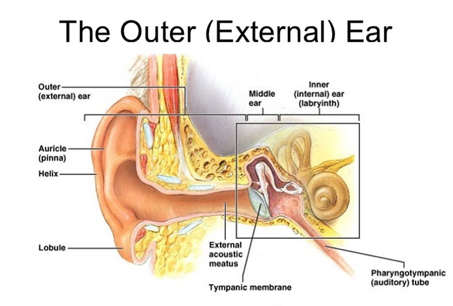
Reading time: 40 minutes Recommended video: Introduction to the ear [21:34] Overview of the structures of the outer ear and auditory tube. External acoustic meatus Meatus acusticus externus 1/4 Synonyms: External auditory meatus, External acoustic pore , show more. The ear is a complex part of an even more complex sensory system.
Ear infections explained Dr Mark McGrath
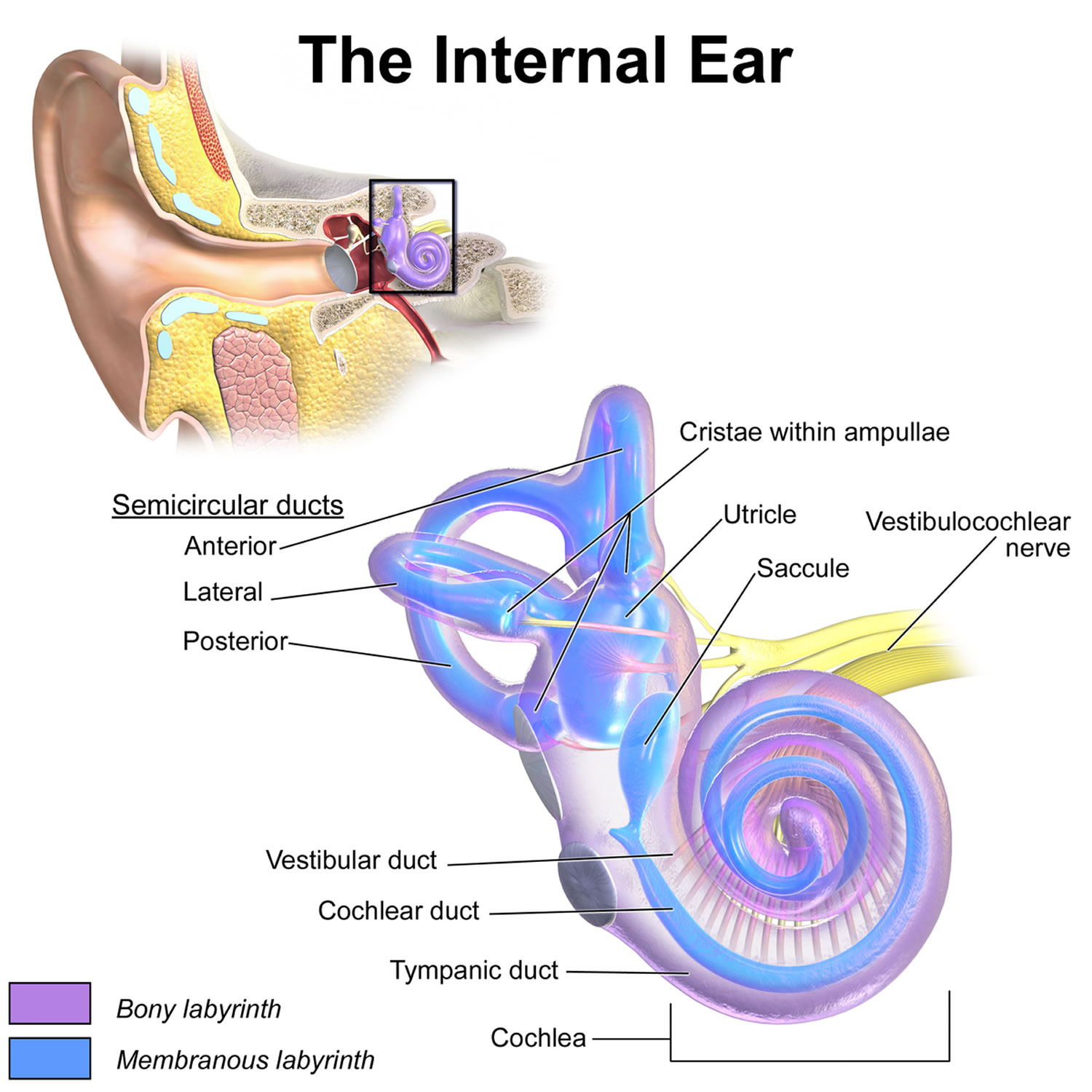
The inner ear, known as the labyrinth, contains two primary structures: the cochlea, responsible for hearing, and the vestibular apparatus, responsible for maintaining balance, stability and spatial orientation. The labyrinth, or inner ear (see figure 1) is encased in bone, called the bony labyrinth. Suspended by fluid (perilymph) within the.
What is conductive hearing loss? Blog of Kiversal
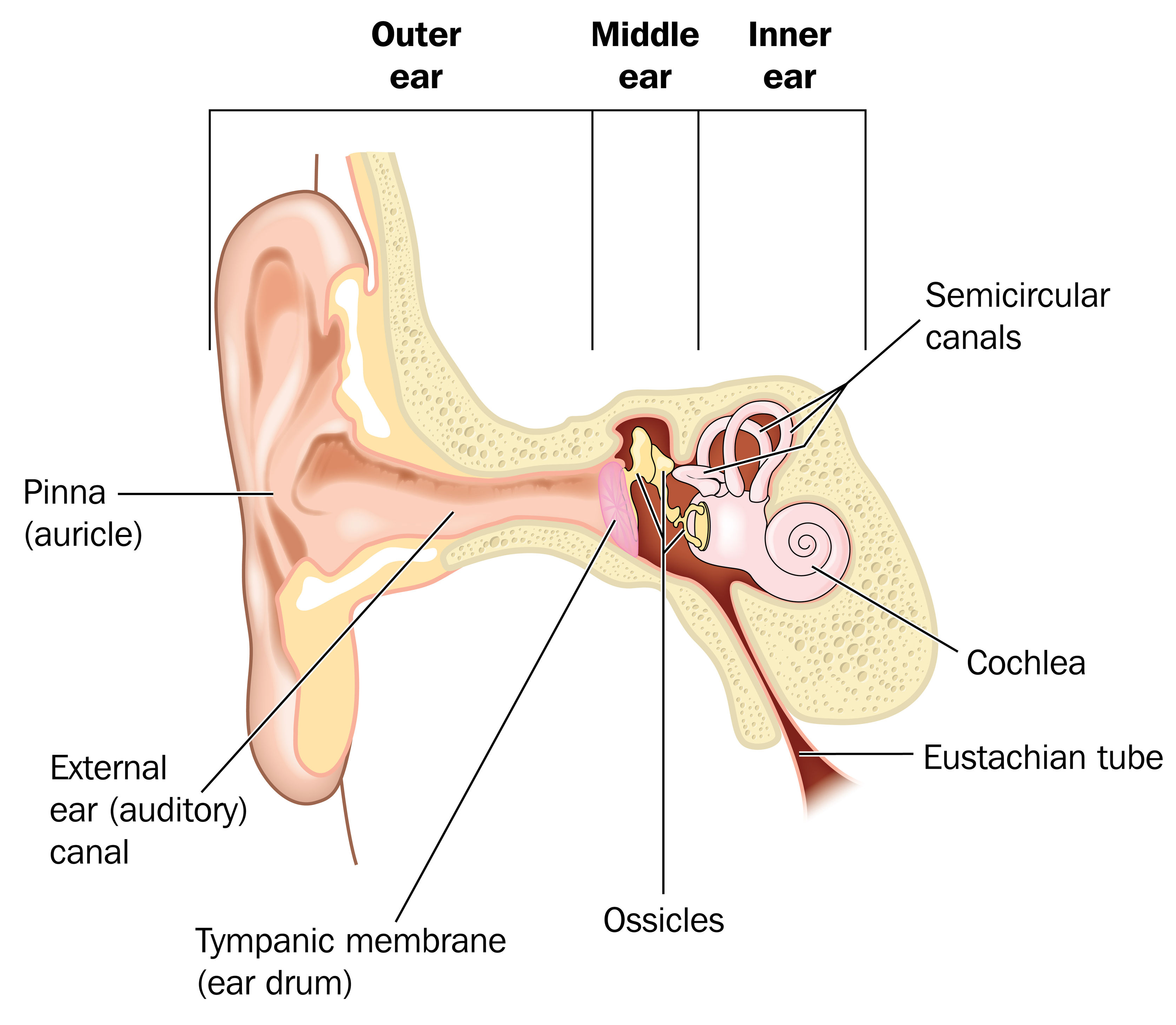
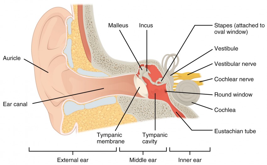
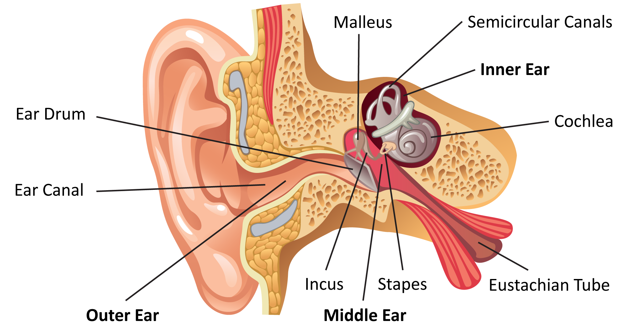
Overview Your outer ear and middle ear are separated by your eardrum, and your inner ear houses the cochlea, vestibular nerve and semicircular canals (fluid-filled spaces involved in balance and hearing). What is the ear? Your ears are organs that detect and analyze sound. Located on each side of your head, they help with hearing and balance.
SPEECH LANGUAGE PATHOLOGY & AUDIOLOGY HEARING DISORDERS OF THE OUTER EAR
Inner and Middle Ear. The cochlea is the most critical component of the inner ear. It is divided into three fluid-filled chambers, called scalae, that spiral around a bony core. The scala media.

Hearing Loss Regenerated in Damaged Mammal Ear The Personal Longevity
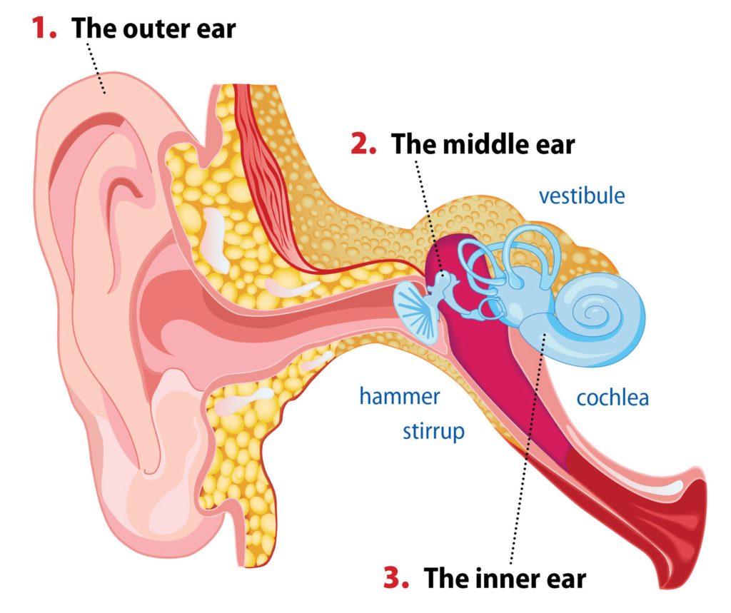
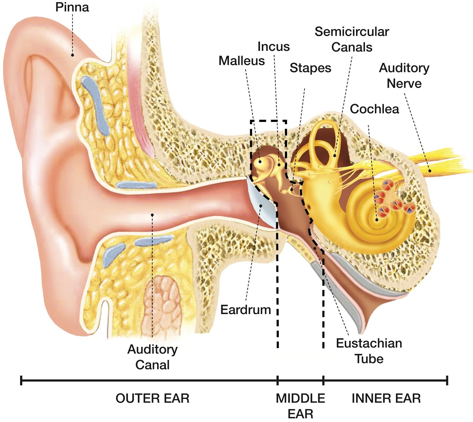
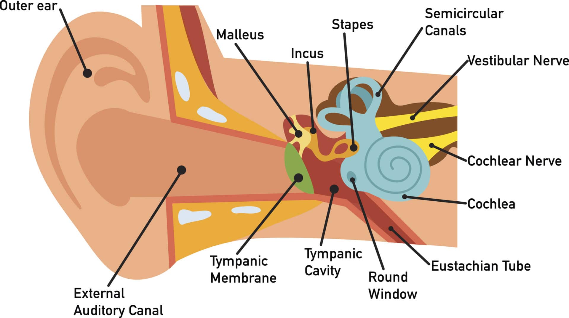
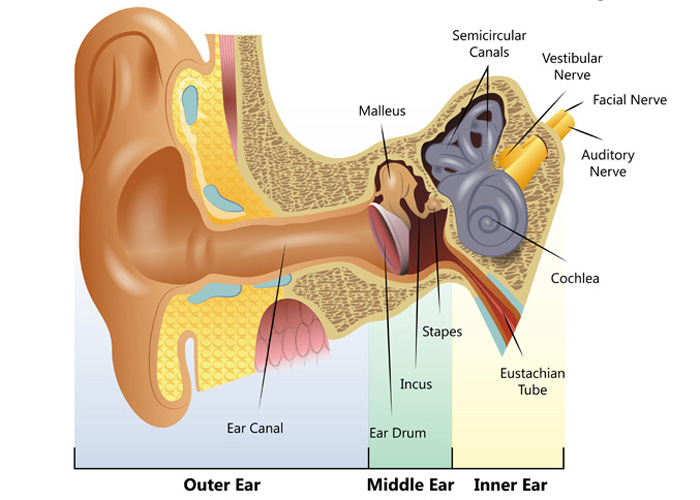
What is the inner ear? What we think of as the "ear" is actually a three-part structure. The outer ear is the part you see and your ear canal. The middle ear is a box-shaped area behind the tympanic membrane (eardrum) that includes the three smallest bones in your body.
Ear Anatomy Causes of Hearing Loss Hearing Aids Audiology
Inner ear anatomy. The outer, middle, and inner ear. The inner ear is at the end of the ear tubes. It sits in a small hole-like cavity in the skull bones on both sides of the head. The inner ear.

Diagram of the anatomy of the ear Ear diagram, Human ear, Ear anatomy
Ear Anatomy, Diagram & Pictures | Body Maps Human body Head Ear Ear The ears are organs that provide two main functions — hearing and balance — that depend on specialized receptors called.

Alila Medical Media Human ear anatomy, labeled diagram. Medical
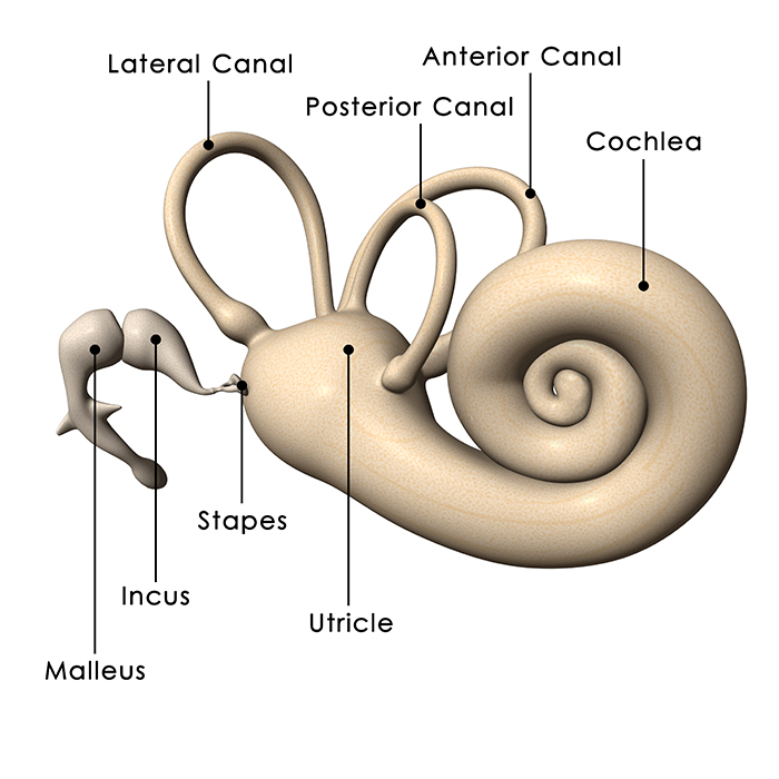
Anatomy. The inner ear is a complex three dimensional shape with semicircular canals, dilations called the utricle and saccule and a spiral portion known as the cochlea. All of these organs are housed inside a bony shell known as the bony labyrinth and this is within the temporal bone. The cochlea is the site where sound is transformed into.
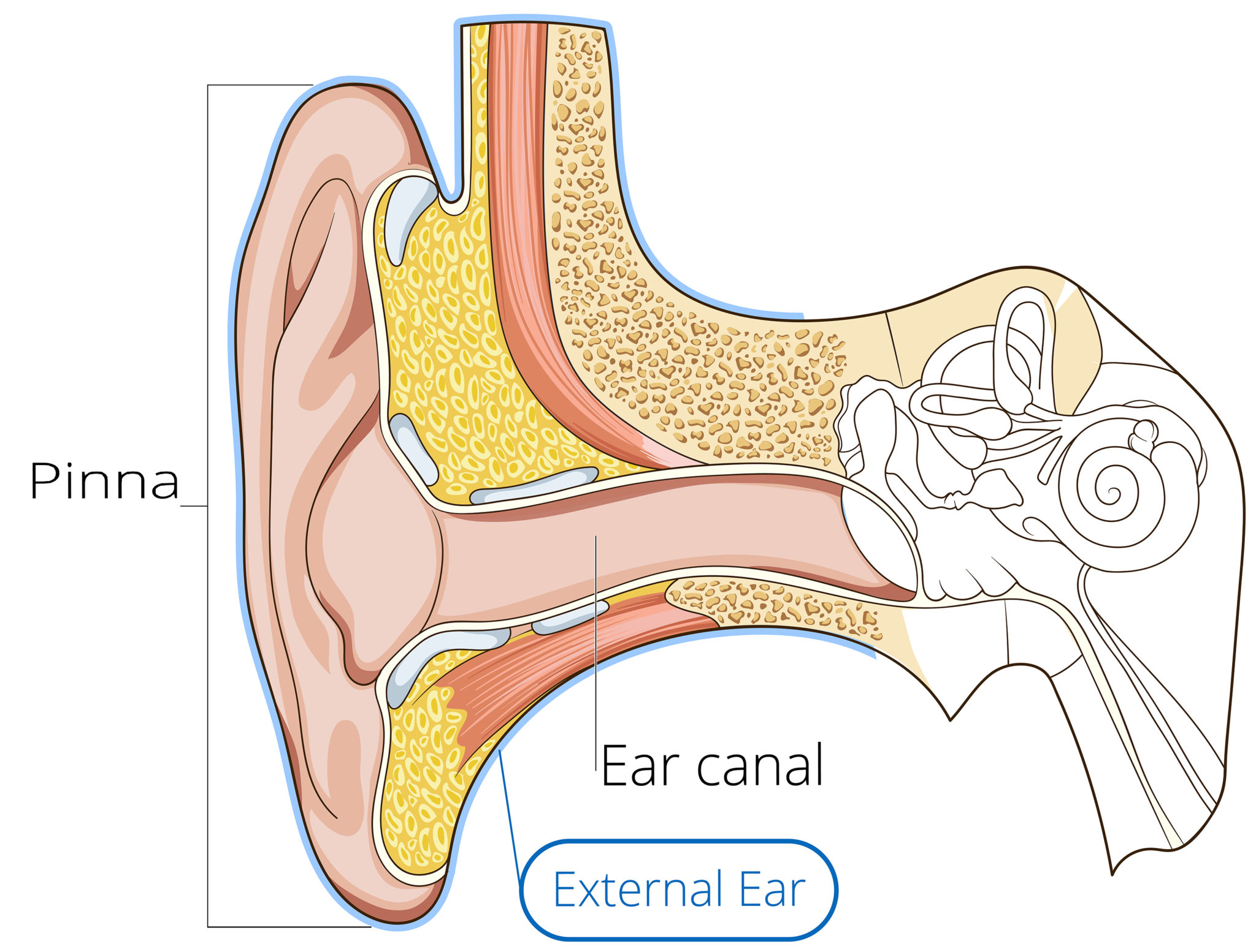
Outer Ear Anatomy Outer Ear Infection & Pain Causes & Treatment
human ear, organ of hearing and equilibrium that detects and analyzes sound by transduction (or the conversion of sound waves into electrochemical impulses) and maintains the sense of balance (equilibrium). Understand the science of hearing and how humans and other mammals perceive sound How humans and other mammals perceive sound.

utricle Google Search EarWaxBuildup Inner ear diagram, Inner ear
The inner ear is embedded within the petrous part of the temporal bone, anterolateral to the posterior cranial fossa, with the medial wall of the middle ear, the promontory, serving as its lateral wall.
We Finally Know Why There's a Bizarre Structure in Our Inner Ears
The fluid inside the cochlea, which is a spiral-shaped, fluid-filled structure in the inner ear, is called perilymph. Perilymph is one of the two types of fluid found in the cochlea, the other being endolymph. These fluids play a crucial role in the process of hearing by transmitting sound vibrations and stimulating the hair cells in the cochlea.
Audition and Somatosensation Anatomy and Physiology I
Ear Anatomy - Inner Ear Next to the middle ear in the bone of the skull is a small compartment which contains the hearing and balance apparatus known as the inner ear. The inner ear has two main parts. The cochlea , which is the hearing portion, and the semicircular canals is the balance portion.
How We Perceive Sound
The inner ear is the innermost part of the ear and consists of the cochlea, auditory nerve, vestibule and semicircular canals. The inner ear is a maze of tubes and passages, referred to as the labyrinth. The inner ear is mainly responsible for balance and detecting sound. The cochlea contains the cells responsible for hearing, the auditory.
How You Hear Northland Audiology
Ear Anatomy - Inner Ear. Ear Anatomy Schematics. Ear Anatomy Images. Chapter 4 - Fluid in the ear. Fluid in the ear Discussion. Fluid in the ear Outline. Middle Ear Ventilation Tubes. Fluid in the ear Images. Chapter 5 - Traveler's Ear.
Inner Ear Problems Causes & Treatment of inner ear Dizziness & Vertigo
Structure and Function. The ear is organized into three different anatomical structures: the outer, middle, and inner ear. The outer ear consists of the pinna, external auditory canal, and tympanic membrane and is responsible for the transmission of sound waves from the external environment. The middle ear is an air-filled space that contains the three ossicles (malleus, incus, and stapes.
Common balance disorders Hearing Link
inner ear, part of the ear that contains organs of the senses of hearing and equilibrium.The bony labyrinth, a cavity in the temporal bone, is divided into three sections: the vestibule, the semicircular canals, and the cochlea.Within the bony labyrinth is a membranous labyrinth, which is also divided into three parts: the semicircular ducts; two saclike structures, the saccule and utricle.